The immune system, which is responsible for the process of antibody biosynthesis, consists of a number of organ systems that include the spleen, thymus and structures in which the formation of three main types of cells occurs, called T and B lymphocytes and macrophages.
The production of antibodies occurs due to the work of B lymphocytes, which on their surface have receptors that bind the antigen. This also includes group T lymphocytes and macrophages. Activation of B lymphocytes occurs as a result of intercellular cooperation, as well as their subsequent transformation into plasma cells.
If the disease develops in your body, you may suffer from slowing down your life and fatigue, constipation, dry skin and so on. will. Often patients also suffer, fall, gain weight and become sensitive to cold. Sometimes a specialist may see a patient with suspected autoimmune disorders such as systemic lupus erythematosus, rheumatoid arthritis, and pernicious anemia.
Patients who plan to include amiodarone, interferon alfa, interleukin 2, lit. This is because the correct values are determined by many factors, including the patient's age, gender, methodology used, and the laboratory where the labeling is made. Therefore the so-called. The range of assignment is determined for a specific sign.
A huge number of plasma cells that have just formed synthesize antibodies that are located on the surface of B lymphocytes, similar to receptors, and then secrete these cells into the blood. The remaining cells become immune memory cells, which enables them to secrete antibodies in the subsequent situation when an antigen enters the blood.
If they rise to antibody levels, Graves' disease or chronic thyroiditis is likely developing. Their presence in the blood may indicate various diseases of the thyroid gland. The test is performed by taking a sample of venous blood, which is sent to a laboratory for analysis.
We think of the “soldiers” that the immune system produces to fight microorganisms that can make the human body sick. But how it works and what it produces, you can find out from this material. Antibodies or proteins called "immunoglobulins" are secreted by type B lymphocytes and are found in body fluids. Typically these proteins react in the presence of antigens or microorganisms that make you sick.
On the surface of each group B lymphocyte there are approximately 100,000 receptors of identical specificity. When an antigen enters the bloodstream, it encounters a receptor called complement. Next, the process of selecting a suitable group B lymphocyte occurs, which, in turn, is transformed into a plasma cell and undergoes multiple divisions, forming a collection of cells.
In other words, antigens are foreign substances that threaten the body and cause the immune system to react. An antigen may produce, after vaccination, for example, an immune response called an "immunogen" needed to produce antibodies.
Antibodies, in turn, can specifically bind to an antigenic determinant or epitope. The rule states that an antibody is capable of recognizing only one antigenic determinant. In other words, if a person gets sick with a certain type of flu, the body reacts through the immune system with the corresponding antigens, producing antibodies that counteract the disease; Antibodies already prepared for influenza will recognize the same type of influenza a second time and will fight against it from the very first signs.
This process was first subjected to a qualitative formulation by P. Ehrlich, and then modified according to the level of scientific progress by F. Burnet, receiving the name “clonal selection theory.” It is worth saying that any of the plasma cell clones is capable of secreting antibodies that have a homogeneous structure. But, due to the simultaneous activation in the bloodstream of several types of group B lymphocytes containing the original antigen, such a response is called “polyclonal”; antibodies began to be called polyclonal.
The immune system can produce other antibodies for other types of diseases. Each time a new type of disease develops, other antibodies are produced and bind to the antigens to destroy them. The immunoglobulins produced by the body are extremely numerous - there are more than 100 different molecular types.
When the body is immunized, an immune response is created; antibodies found in the blood, in particular. All antibodies have the same basic structure - they are glycoprotein molecules produced by activated plasma cells or B lymphocytes. There are two types of immunoglobulins according to their structure.
Animal serum that contains antibodies specific to this antigen is called antiserum. There is often an indication of which antigen the serum was produced against. For example, when talking about serum that contains antibodies to the red blood cells of the human body, we mean that the response of human blood to the introduction of rabbit red blood cells is expressed in the production of specific antibodies.
Antibodies have a greater ability to fight antigens if they bind strongly to the latter. Classification of immunoglobulins. Immunoglobulins vary depending on how these antibodies bind to antigens. What does each immunoglobulin do?
Helps neutralize bacteria; therefore, if they are missing from the body, there is a risk of bacterial infections. Increased level can be found if there is a predisposition to multiple sclerosis or hepatitis. They are the main antibodies and are found in tears, saliva, respiratory, urogenital or gastrointestinal secretions.
If the antigen cells are polyvalent, speaking of a protein, for example, then the formation of antibodies occurs in the bloodstream, which are intended separately for a specific determinant. This significantly complicates the process of antibody formation. The composition of each antibody depends on the species of animal, including the stage of the process in the immune system.
This is a barrier against bacteria. Generally speaking, antibodies react to recognized antigens in the body, antigens to which they bind and which they destroy. It should be noted that a lower amount of immunoglobulins or an increased amount are signs of the disease.
Everything we publish. Below are detailed explanations of gluten intolerance tests to help you better understand the celiac disease diagnosis process. First of all, before you do any blood tests, you should not be removing gluten from your diet, so the tests are correct. Antibodies are produced by the immune system in response to substances that the body perceives as dangerous to it. The immune response that the body produces is caused by exposure to gluten in the diet. If there is no source of gluten in the diet, then there is no body response that can be measured. If you had it for a short period of time, such as a week, then the response might be reduced, but the difference would be negligible because the body did not have time to respond to the change. On the other hand, if you have been on a gluten-free diet for a longer period of time, there is a risk of getting the wrong results. You need to eat at least 2 slices of bread per day for 6 weeks to get accurate results. There are basically four tests that can be performed to help diagnose celiac disease. Please note that all laboratory tests, regardless of type, are presented as diagnostic tools. They should not be used alone as a basis for diagnosis, but should be considered in close connection with the patient's physical examination as well as the present symptoms. In our bodies there is a family of closely related, although not identical, proteins that are capable of acting as antibodies.
Everything described above affects the process of antibody heterogeneity, which is caused by certain difficulties in the study of their structure, including the production of universal antiserum compositions. The study of hybridization by Köhler and Milstein opened up new routes that made it possible to obtain antibodies. The essence of this approach by scientists is that the body of an immunized animal secretes lymphocytes, which subsequently merge with cells called “myeloma.” The cells that are formed are called hybrids.
How do we interpret gluten intolerance?
They are called immunoglobulins. Thus, there are five main types of immunoglobulins. Each of them has different functions in our systems. It produces antibodies associated with most hypersensitivity responses. These names refer to the type of protein that carries the antibody. Thank you for subscribing to the newsletter!
These antibodies are found in 100% of patients with active celiac disease. Tissue transglutaminase has been identified as a major autoantigen in celiac disease. In this test, the reaction is measured using an instrument that calculates the amount of light of a given wavelength that is absorbed by a solution and finally outputs a numerical result. There is no human error because there is no human judgment.
Hybrid cells are special in that they can multiply and produce antibodies in artificially created conditions outside the human body. Thanks to special methods, one cell is isolated - a hybrid, which will be capable of secreting a huge amount of antibodies of only a specific species - monoclonal antibodies.
How our immune system works
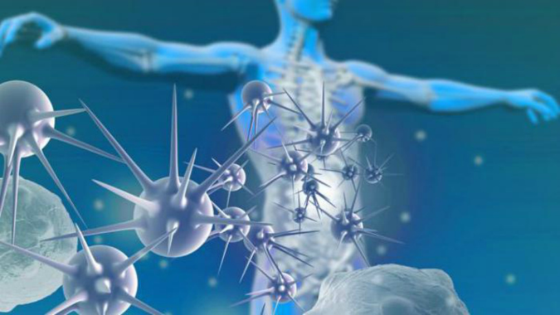
The very first cells that a virus enters the body will encounter are macrophages. These cells are constantly ready to protect our body, and act as cleansers in the blood. They are in constant dynamics and are ready to repel any attack of viruses on the body. The peculiarity of macrophages is the recognition of microbes and viruses that differ from the cells of our body. The structure and functional features allow these cells to instantly destroy foreign agents, and then continue to move throughout the body.
But the number of macrophages is not always able to defeat a huge number of harmful agents, as evidenced by a sharp increase in body temperature. This is explained by the production of a special substance - pyrogen, which reaches the brain cells and then stimulates the thermoregulatory center. As soon as the pyrogen reaches this center, the body begins to suffer from fever, weakness, and malaise. During this period, it is recommended to be in bed, as the immune forces should increase and recover.
In the process of macrophages fighting enemy foreign cells, lymphocytes of group T and B join the process. Once the macrophage has caught and destroyed the virus, it becomes the target point for reconnaissance cells, which are called information T cells. This is a fairly active set of cells that reads and recognizes all the data that viruses carry, and then sends signals throughout the body about the need to activate immune defense.
As soon as the cells immune system received a signal from T cells, the warning is carried to every cell of the systems and organs of the whole body. As a result of this process, a biologically active molecule called lymphokine is released, which coordinates the readiness of immune cells. Thus, group B lymphocytes begin to produce antibodies, which will neutralize and destroy foreign organisms.
Lymphocytes of group B and T - cells carry out their activities together, as evidenced by the lightning-fast production of antibodies, of which T - cells can produce about several thousand in 1 second. Thus, T and B cells are in close information connection, which has a beneficial effect in the fight against foreign viruses.
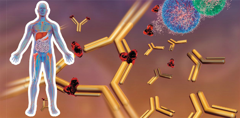
Antibodies have a three-dimensional protein structure, which makes it possible to accurately recognize which part of the bacterium should be blocked, and subsequently broken. This makes it possible to neutralize antibodies that are produced by group B cells and to accurately identify the weak points of bacteria, which becomes known thanks to the work of T cells.
As soon as our body encounters a new disease, namely group B cells begin to produce antibodies that are fatal to the virus of this disease. The process of producing antibodies by our body does not have errors. The information delivered by helper cells does not limit the functions of these cells. There is also a process of involvement of killer T viruses in the fight against viruses. The entry of bacteria and viruses is a serious process, which requires that harmful cells will hide from antibodies in healthy cells, which complicates the task, since antibodies cannot enter inner part healthy cell.
It happens that antibodies cannot recognize the presence of a virus in a healthy cell, since nothing indicates this. In this case, T cells come to the rescue. Their capabilities make it possible to recognize the presence of a pathological process in a cell that, by external parameters, looks healthy. T cells are able to detect a virus in a healthy cell and destroy the healthy cell along with the foreign agent in it. Along with this, macrophages also act, which are responsible for the process of destroying harmful cells and cleansing the body as a whole. As soon as the virus is defeated, calming cells called T-suppressors begin their work. They calm the body, which leads to the normalization of all its processes.
Our body's database
Some cells of group B are predetermined for a long existence and they are also designed to store records of the structures of viruses that have ever entered and are rampant in the body. This feature of the cells in the body is necessary, because in the event of repeated infection of the body by the virus, the cells of the immune system quickly recognize the harmful agent and destroy it as quickly as possible.
Repeated entry into the body of the same virus gives a signal to immune cells to begin producing antibodies against this virus in order to eliminate it. In addition, in such a situation, a person does not suffer from the symptoms of the disease. Cells immune memory have existed much longer than others.
As soon as the moment of the end of their existence comes, they undergo division. At the same time, very interesting feature these cells have - they transmit the entire amount of information that has accumulated over the course of existence about diseases that have ever affected the body to the newly formed cells. This advantage of immune cells is an infectious disease that was transmitted by a person to early age, will not be able to infect the body, since it has immunity specifically to this disease.
Our body's ability to fight harmful bacteria is amazing. After all, small cells are able to resist serious pathogens that can destroy our immunity and lead to serious consequences of the disease. The work of immune cells is colossal, since if the defense function were under human control, we would probably make many mistakes, which would have consequences for the immune system and the entire body as a whole.
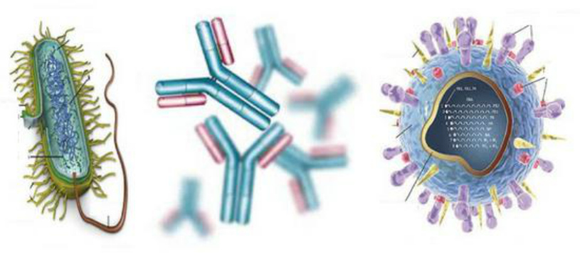
Antibodies- specific proteins of gamma-globulin nature, formed in the body in response to antigenic stimulation and capable of specifically interacting with the antigen (in vivo, in vitro). In accordance with the international classification, the totality of serum proteins that have the properties of antibodies is called immunoglobulins.
The uniqueness of antibodies lies in the fact that they are able to specifically interact only with the antigen that caused their formation.
Immunoglobulins (Ig) are divided into three groups depending on location:
Serum (in the blood);
Secretory (in secretions - the contents of the gastrointestinal tract, lacrimal secretion, saliva, especially in breast milk) provide local immunity(mucosal immunity);
Superficial (on the surface of immunocompetent cells, especially B-lymphocytes).
Any antibody molecule has a similar structure (Y-shaped) and consists of two heavy (H) and two light (L) chains linked by disulfide bridges. Each antibody molecule has two identical antigen-binding fragments Fab (fragment antigen binding), which determine antibody specificity, and one Fc (fragment constant) fragment, which does not bind antigen, but has effector biological functions. It interacts with “its” receptor in the membrane of various types of cells (macrophage, mast cell, neutrophil).
The terminal regions of the light and heavy chains of the immunoglobulin molecule are variable in composition (amino acid sequences) and are designated as VL and VH regions. They contain hypervariable regions that determine the structure active site of antibodies (antigen-binding center or paratope). It is with it that the antigenic determinant (epitope) of the antigen interacts. The antigen-binding center of antibodies is complementary to the antigen epitope according to the “key-lock” principle and is formed by the hypervariable regions of the L- and H-chains. The antibody will bind to the antigen (the key will fit into the lock) only if the determinant group of the antigen fits completely into the gap of the active center of the antibodies.
Light and heavy chains consist of separate blocks - domains. In light (L) chains there are two domains - one variable (V) and one constant (C), in heavy (H) chains - one V and 3 or 4 (depending on the immunoglobulin class) C domains.
There are two types of light chains - kappa and lambda, they are found in different proportions in different (all) classes of immunoglobulins.
Revealed five classes of heavy chains - alpha (with two subclasses), gamma (with four subclasses), exilon, mu and delta. According to the designation of the heavy chain, the class of immunoglobulin molecules is also designated - A, G, E, M and D.
It is the constant regions of heavy chains, differing in amino acid composition in different classes of immunoglobulins, that ultimately determine the specific properties of immunoglobulins of each class.
There are five known classes of immunoglobulins, differing in the structure of heavy chains, molecular weight, physicochemical and biological characteristics: IgG, IgM, IgA, IgE, IgD. There are 4 subclasses of IgG (IgG1, IgG2, IgG3, IgG4), and two subclasses of IgA (IgA1, IgA2).
The structural unit of antibodies is monomer, consisting of two light and two heavy chains. The monomers are IgG, IgA (serum), IgD and IgE. IgM- pentamer(polymeric Ig). Polymer immunoglobulins have an additional j (joint) polypeptide chain that unites (polymerizes) individual subunits (composed of the IgM pentamer, di- and trimer of secretory IgA).
Basic biological characteristics of antibodies.
1. Specificity- the ability to interact with a specific (own) antigen (correspondence between the epitope of the antigen and the active center of the antibodies).
2 . Valence- the number of active centers capable of reacting with the antigen (this is due to the molecular organization - mono- or polymer). Immunoglobulins can be divalent(IgG) or polyvalent(IgM pentamer has 10 active sites). Bi- or more valent antibodies emerge full antibodies. Incomplete antibodies have only one active center involved in interaction with the antigen (blocking effect on immunological reactions, for example, on agglutination tests). They are detected in the Coombs antiglobulin test, the reaction of inhibition of complement fixation.
3. Affinity - the strength of the connection between the antigen epitope and the active center of antibodies depends on their spatial correspondence.
4. Avidity - an integral characteristic of the strength of connection between antigen and antibodies, taking into account the interaction of all active centers of antibodies with epitopes. Since antigens are often multivalent, communication between individual antigen molecules is carried out by several antibodies.
5. Heterogeneity - due to the antigenic properties of antibodies, the presence of three types of antigenic determinants:
- isotypic- antibodies belong to a certain class of immunoglobulins;
- allotypic- caused by allelic differences in immunoglobulins encoded by the corresponding alleles of the Ig gene;
- idiotypal- reflect individual characteristics immunoglobulin, determined by the characteristics of the active centers of antibody molecules. Even when antibodies to a particular antigen belong to the same class, subclass, or even allotype, they are characterized by specific differences from each other ( idiotic). This depends on the structural features of the V-sections of the H- and L-chains, the set various options their amino acid sequences.
The concept of polyclonal and monoclonal antibodies will be given in the following sections.
Characteristics of the main classes of immunoglobulins.
Ig G. Monomers include four subclasses. Concentration in the blood is from 8 to 17 g/l, half-life is about 3-4 weeks. This is the main class of immunoglobulins that protect the body from bacteria, toxins and viruses. The largest quantities of IgG antibodies are produced at the stage of recovery after an infectious disease (late or 7S antibodies), during the secondary immune response. IgG1 and IgG4 specifically (through Fab fragments) bind pathogens ( opsonization), thanks to the Fc fragments of IgG, they interact with the Fc receptors of phagocytes, promoting phagocytosis and lysis of microorganisms. IgG is capable of neutralizing bacterial exotoxins and fixing complement. Only IgG is able to be transported across the placenta from mother to fetus (pass through the placental barrier) and provide protection by maternal antibodies to the fetus and newborn. Unlike IgM antibodies, IgG antibodies belong to the late category - they appear later and are detected in the blood for a longer time.
IgM. The molecule of this immunoglobulin is a polymeric Ig of five subunits connected by disulfide bonds and an additional J-chain, and has 10 antigen-binding centers. Phylogenetically, this is the most ancient immunoglobulin. IgM is the earliest class of antibodies formed when an antigen first enters the body. The presence of IgM antibodies to the corresponding pathogen indicates a fresh infection (current infectious process). Antibodies to antigens of gram-negative bacteria, flagellar antigens - predominantly IgM antibodies. IgM is the main class of immunoglobulins synthesized in newborns and infants. IgM in newborns is an indicator of intrauterine infection (rubella, CMV, toxoplasmosis and other intrauterine infections), since maternal IgM does not pass through the placenta. The concentration of IgM in the blood is lower than IgG - 0.5-2.0 g/l, half-life is about a week. IgM is capable of agglutinating bacteria, neutralizing viruses, activating complement, activating phagocytosis, and binding endotoxins of gram-negative bacteria. IgM has greater avidity than IgG (10 active centers), affinity (affinity for antigen) is less than that of IgG.
IgA. There are serum IgA (monomer) and secretory IgA (IgAs). Serum IgA is 1.4-4.2 g/l. Secretory IgAs are found in saliva, digestive juices, secretions of the nasal mucosa, and colostrum. They are the first line of defense for mucous membranes, providing their local immunity. IgAs consist of an Ig monomer, a J chain, and a glycoprotein (secretory component). There are two isotypes: IgA1 predominates in serum, subclass IgA2 in extravascular secretions.
The secretory component is produced by epithelial cells of the mucous membranes and attaches to the IgA molecule as it passes through the epithelial cells. The secretory component increases the resistance of IgAs molecules to the action of proteolytic enzymes. The main role of IgA is to provide local immunity to the mucous membranes. They prevent bacteria from attaching to mucous membranes, ensure the transport of polymeric immune complexes with IgA, neutralize enterotoxin, and activate phagocytosis and the complement system.
IgE. It is a monomer and is found in low concentrations in blood serum. The main role is that with its Fc fragments it attaches to mast cells (mast cells) and basophils and mediates immediate hypersensitivity reactions. IgE refers to “allergy antibodies” - reagins. The level of IgE increases in allergic conditions and helminthiasis. The antigen-binding Fab fragments of the IgE molecule specifically interact with the antigen (allergen), the formed immune complex interacts with the receptors of the Fc fragments of IgE embedded in the cell membrane of the basophil or mast cell. This is a signal for the release of histamine and other biologically active substances and the development of an acute allergic reaction.
IgD. IgD monomers are found on the surface of developing B lymphocytes and are found in extremely low concentrations in serum. Their biological role has not been clearly established. It is believed that IgD is involved in the differentiation of B cells, contributes to the development of an anti-idiotypic response, and is involved in autoimmune processes.
To determine the concentrations of immunoglobulins of individual classes, several methods are used, most often method of radial immunodiffusion in gel (according to Mancini) - a type of precipitation reaction and ELISA.
Determination of antibodies of various classes is important for the diagnosis of infectious diseases. Detection of antibodies to antigens of microorganisms in blood serum is an important criterion when making a diagnosis - serological diagnostic method. Antibodies of the IgM class appear in the acute period of the disease and disappear relatively quickly, antibodies of the IgG class are detected in more late dates and persist for a longer period (sometimes for years) in the blood serum of those who have recovered from the disease; in this case they are called anamnestic antibodies.
The concepts are distinguished: antibody titer, diagnostic titer, studies of paired sera. The most important is the detection of IgM antibodies and a fourfold increase in antibody titers (or seroconversion- antibodies are detected in the second sample with negative results with the first blood serum) during the study doubles- taken in the dynamics of the infectious process at intervals of several days-weeks samples
Reactions of interaction of antibodies with pathogens and their antigens ( reaction “ antigen-antibody”) manifests itself in the form of a number of phenomena - agglutination, precipitation, neutralization, lysis, complement fixation, opsonization, cytotoxicity and can be identified by various serological reactions.
Dynamics of antibody production. Primary and secondary immune response.
The primary response is upon initial contact with the pathogen (antigen), the secondary response is upon repeated contact. Main differences:
Duration of the latent period (longer during the primary period);
The rate of growth of antibodies (faster in secondary);
The amount of antibodies synthesized (more with repeated contact);
The sequence of synthesis of antibodies of different classes (in the primary, IgM predominates for a longer time, in the secondary, IgG antibodies are quickly synthesized and predominate).
The secondary immune response is due to the formation immune memory cells. An example of a secondary immune response is an encounter with a pathogen after vaccination.
The role of antibodies in the formation of immunity.
Antibodies are important in the formation acquired post-infectious and post-vaccination immunity.
1. By binding to toxins, antibodies neutralize them, providing antitoxic immunity.
2. By blocking virus receptors, antibodies prevent the adsorption of viruses on cells and participate in antiviral immunity.
3. The antigen-antibody complex triggers the classical pathway of complement activation with its effector functions (bacterial lysis, opsonization, inflammation, stimulation of macrophages).
4. Antibodies take part in the opsonization of bacteria, promoting more efficient phagocytosis.
5. Antibodies promote the excretion of soluble antigens from the body (with urine, bile) in the form of circulating immune complexes.
IgG plays the largest role in antitoxic immunity, IgM - in antimicrobial immunity (phagocytosis of corpuscular antigens), especially against gram-negative bacteria, IgA - in antiviral immunity (neutralization of viruses), IgAs - in local immunity of mucous membranes, IgE - in immediate hypersensitivity reactions .


